Сурет:Virus Replication.svg

Түпнұсқа файл (SVG файлы, кесімді 462 × 426 (пиксел) нүкте, файл өлшемі: 205 КБ)
| Бұл файл Wikimedia Commons? жобасынан, сондықтан басқа жобаларда да қолдануы мүмкін. Commons ашық лицензиялы медиа файл қоры. Сіз жобаға көмектесе аласыз. |
Ортаққордан қарау |
Түйін
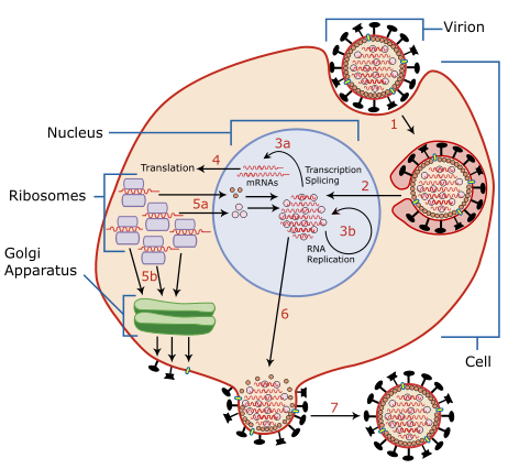
| Сипаттамасы | A diagram of influenza viral cell invasion and replication. |
| Күні | |
| Көзі | Redrawn from w:Image:Virusreplication.png using Adobe Illustrator. |
| Авторы | User:YK Times |
| Басқа нұсқалары |
 |


|
This SVG file contains embedded text that can be translated into your language, using any capable SVG editor, text editor or the SVG Translate tool. For more information see: About translating SVG files. |
Description from Scheme of Influenza A virus replication (NCBI): "A virion attaches to the host cell membrane via HA and enters the cytoplasm by receptor-mediated endocytosis (STEP 1), thereby forming an endosome. A cellular trypsin-like enzyme cleaves HA into products HA1 and HA2 (not shown). HA2 promotes fusion of the virus envelope and the endosome membranes. A minor virus envelope protein M2 acts as a ion channel thereby making the inside of the virion more acidic. As a result, the major envelope protein M1 dissociates from the nucleocapsid and vRNPs are translocated into the nucleus (STEP 2) via interaction between NP and cellular transport machinery. In the nucleus, the viral polymerase complexes transcribe (STEP 3a) and replicate (STEP 3b) the vRNAs. Newly synthesized mRNAs migrate to cytoplasm (STEP 4) where they are translated. Posttranslational processing of HA, NA, and M2 includes transportation via Golgi apparatus to the cell membrane (STEP 5b). NP, M1, NS1 (nonstructural regulatory protein - not shown) and NEP (nuclear export protein, a minor virion component - not shown) move to the nucleus (STEP 5a) where they bind freshly synthesized copies of vRNAs. The newly formed nucleocapsids migrate into the cytoplasm in a NEP-dependent process and eventually interact via M1 with a region of the cell membrane where HA, NA and M2 have been inserted (STEP 6). Then the newly synthesized virions bud from infected cell (STEP 7). NA destroys the sialic acid moiety of cellular receptors, thereby releasing the progeny virions."
Лицензиялау

|
Бұл файлды GNU Free Documentation License лицензиясының 1.2 нұсқасы бойынша немесе ескі Ашық бағдарламалық жасақтаушы қорымен жарияланған нұсқасының шарттарына сәйкес көшірмесін алуға, таратуға және/немесе өзгертуге болады. Лицензия көшірмесі GNU Free Documentation License деп аталынған бөлімде көрсетілген. |
| Бұл файл Creative Commons Attribution-Share Alike 3.0 Unported лицензиясы бойынша қолжетімді. | ||
| ||
| Лицензияландырудың бұл қасиеті осы файлға GFDL лицензиясының жаңартылуының бір бөлігі ретінде енгізілген. |
- Сіз келесі әрекеттерге еркінсіз:
- бөлісу – туындыны көшіру, тарату және тапсыру
- мазмұнын өзгерту – туындыны бейімдеу
- Келесі ережелерді сақтағанда:
- атрибуция – Авторлықты белгілеп, лицензияға сілтеме беріп, өзгеріс жасалғанын анықтауыңыз керек. Сіз мұны кез келген орынды жолмен істей аласыз, бірақ лицензиар сізді немесе қолдануыңызды мақұлдайтындай емес.
- бірдей шарттарда тарату – Материалды араластырсаңыз, түрлендірсеңіз немесе құрастырсаңыз, үлестеріңізді түпнұсқамен бірдей бірдей немесе үйлесімді лицензия бойынша таратуыңыз керек.
| Annotations | This image is annotated: View the annotations at Commons |
Файл тарихы
Файл сол кезде қалай көрінгенін көру үшін күн/уақыт дегенге басыңыз.
| Күн/Уақыт | Нобай | Өлшемдер | Қатысушы | Пікір | |
|---|---|---|---|---|---|
| қазіргі | 08:54, 2007 ж. наурыздың 6 | | 462 × 426 (205 КБ) | YK Times | {{Information |Description=A diagram of influenza viral cell invasion and replication. |Source=Redrawn from w:Image:Virusreplication.png using Adobe Illustrator. |Date=March 5, 2007 |Author= User:YK Times |Permission= |other_versions=[[:w:Image:V |
Файл қолданылуы
Бұл файлды мына бет қолданады:
Глобалды файл қолданылуы
Бұл файл келесі басқа уикилерде қолданылады:
- bg.wikipedia.org жобасында қолданылуы
- bn.wikipedia.org жобасында қолданылуы
- br.wikipedia.org жобасында қолданылуы
- ca.wikipedia.org жобасында қолданылуы
- cs.wikipedia.org жобасында қолданылуы
- da.wikipedia.org жобасында қолданылуы
- de.wikipedia.org жобасында қолданылуы
- el.wikipedia.org жобасында қолданылуы
- en.wikipedia.org жобасында қолданылуы
- Orthomyxoviridae
- Amantadine
- Viral replication
- Viral life cycle
- Viral entry
- File:Virusreplication.png
- User:YK Times/Graphic Lab/examples
- Wikipedia:Graphics Lab/Images to improve/Archive/Mar 2007
- Viral shedding
- Template:Influenza virus life cycle
- Viral neuraminidase
- Tilapia tilapinevirus
- User:Zoe.gum/sandbox
- Wikipedia talk:WikiProject Viruses/Archive 4
- User:Anicm1/sandbox
- en.wikibooks.org жобасында қолданылуы
- es.wikipedia.org жобасында қолданылуы
- fa.wikipedia.org жобасында қолданылуы
- fr.wikipedia.org жобасында қолданылуы
- hy.wikipedia.org жобасында қолданылуы
- id.wikipedia.org жобасында қолданылуы
- it.wikipedia.org жобасында қолданылуы
- ja.wikipedia.org жобасында қолданылуы
- ko.wikipedia.org жобасында қолданылуы
Бұл файлдың глобалды қолданылуын көбірек көру.